
Vision Refractive Suite at AKEH
Experience the world’s most advanced topography-guided laser system. Tailored to your unique eye “fingerprint” for high-definition clarity.
Stop seeing through lenses and start seeing the world in HD. With Alcon Contura Vision at Dr. Anil Kulkarni Eye Hospital, we don’t just remove your glasses—we upgrade your vision. Experience life in high definition.
What is Contura Vision?
While traditional LASIK corrects your glasses prescription, Contura Vision (Topography-Guided LASIK) goes a step further. It uses the Alcon WaveLight® Refractive Suite to map 22,000 unique elevation points on your cornea. Think of it as a custom-tailored suit versus one off-the-rack; it’s designed specifically for the unique contours of your eyes.
Why Choose the Alcon Refractive Suite at AKEH?
- Precision Beyond Comparison: Maps 22,000 points (standard LASIK only maps 200).
- Superior Visual Outcomes: Many patients achieve vision better than 6/6, with enhanced contrast and color.
- Reduced Side Effects: Significantly minimizes night-driving issues, glares, and halos compared to older technologies.
- The AKEH Trust: Combined with our 45 years of clinical expertise and NABH accreditation, you are in the safest hands in Western Maharashtra.
The Procedure – Fast, Painless, Bladeless
- Digital Mapping : We create a 3D “Topographic Map” of your eye.
- Personalized Planning : The Alcon suite calculates a treatment profile unique to you.
- Laser Correction : The procedure takes less than 10-15 minutes, is completely painless (using numbing drops), and offers a recovery time of just 24-48 hours.
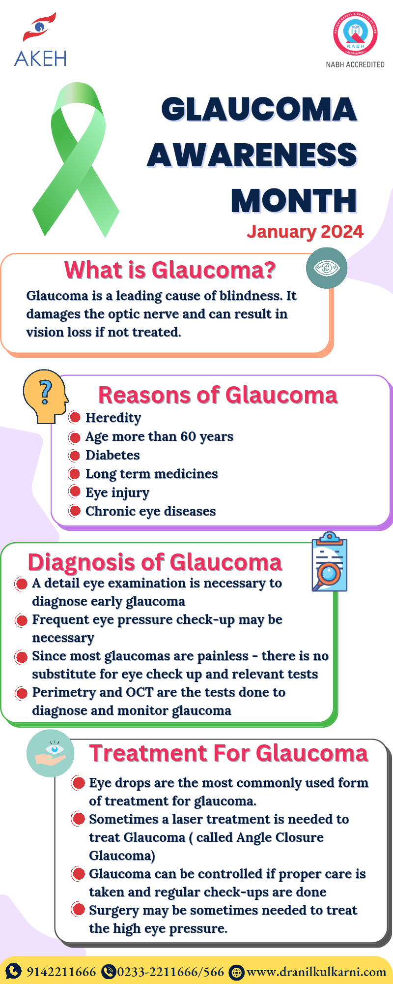
What is Glaucoma?
Glaucoma is a common eye condition in which vision is lost because of damage to the optic nerve. The optic nerve carries information about vision from the eye to the brain. In most cases, the optic nerve is damaged when the pressure of fluid inside the front part of the eye rises. However, glaucoma-related eye damage can occur even when the fluid pressure is normal. An untreated Glaucoma can result in a gradual, irreversible loss of vision and eventual blindness.
Types of Glaucoma
In the most common form of glaucoma, called Open Angle Glaucoma: pressure tends to rise slowly over time. Patients present late in the disease. Gradual loss of vision is only the complaint. A less common form of the disease, called Closed Angle Glaucoma, develops suddenly and usually causes eye pain and redness. In addition, there are rarer forms of glaucoma. They may be related to eye defects that develop before birth (congenital glaucoma) or to eye injuries, eye tumors, or medical problems such as diabetes. In some cases, medications, such as corticosteroids, can also trigger glaucoma.
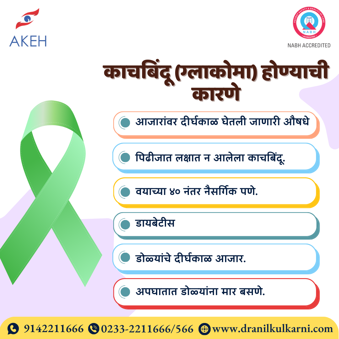
Risk Factors
- Age of 40 and older
- Family history of glaucoma
- Suffer from high plus or minus number
- Have diabetes, hypertension, or heart disease
- Have high intraocular pressure (IOP)
- Experienced eye pain/swelling after previous surgery
- Injury to the eye
- Long duration of medications in the eye or taken orally (especially steroids)
Symptoms
Although open angle glaucoma and closed angle glaucoma both can cause blindness, their symptoms are very different.
Open Angle Glaucoma: In this form of glaucoma, vision is lost painlessly and so gradually that most people do not realize they have a problem until substantial damage has occurred. Peripheral vision (at the edges) is usually lost first. As larger areas of your peripheral vision fade, you may develop tunnel vision—vision that has narrowed so you see only what is directly in front of you, like looking through a railroad tunnel. If glaucoma is not treated, even this narrowed vision disappears into blindness. Once gone, areas of lost vision cannot be restored.
Closed Angle Glaucoma: Symptoms of acute glaucoma occur suddenly and can include blurred vision, pain and redness in the eye, severe headache, halos around lights at night, a haziness in the cornea (the clear front portion of the eye in front of the pupil), nausea and vomiting, and extreme weakness. Glaucoma is a lifelong illness, but proper treatment can prevent loss of vision.
Recommendations
Everyone under 40 years should have an eye examination at least once in four years and after 40 at least once in 2 years. Everyone under 40 years with one of the aforementioned risk factors should have an eye examination at least once in 2 years and after 40 at least once a year.
Diagnosis
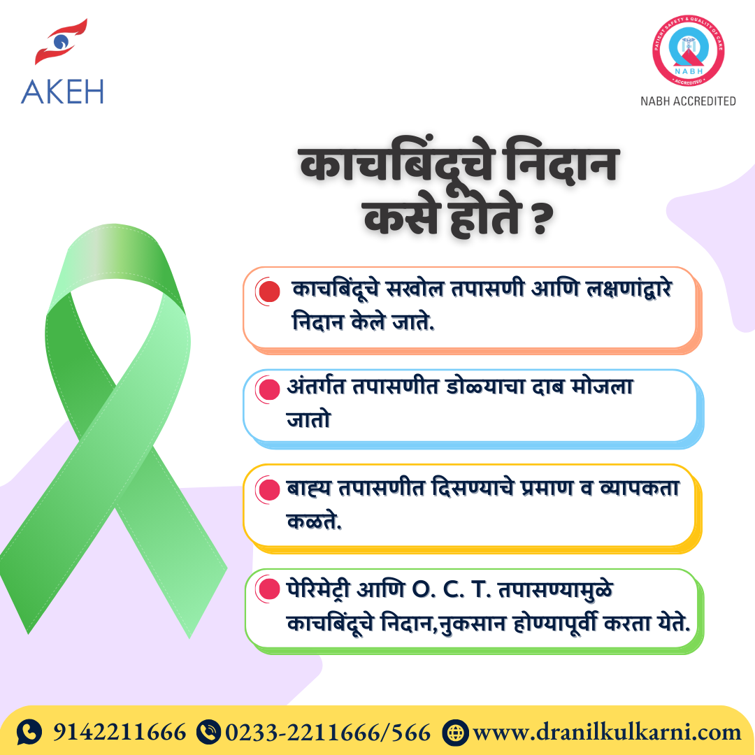
In most cases, Open Angle Glaucoma is diagnosed by a doctor during a routine eye examination. If glaucoma is suspected, your doctor will confirm the diagnosis with one or more additional tests:
- Tonometry: measures the pressure within the eye.
- Gonioscopy: evaluation of the Anterior chamber angle to check if the angle is open or closed.
- Anterior segment OCT: These recent imaging techniques give a good objective evidence of the abnormalities of the angle.
- Visual-field testing: is the best way to find early signs of loss of peripheral vision. Most often, visual fields are checked using an automated machine.
- OCT: This technique uses a LASER beam to actually measure the thickness of the nerve fibers in the retina. Glaucoma causes loss of these nerve fibers.
Treatment
- Eye drops to lower the pressure: Treatment of Open Angle Glaucoma usually begins with prescription eye drops. These medicines lower pressure inside the eye.
- Laser procedure: Treatment of Closed Angle Glaucoma usually begins with a laser procedure. Laser procedure may not lower eye pressure to acceptable levels. You may need to start glaucoma eye drops after laser.
- Conventional eye surgery: If both medication and laser surgery are unsuccessful, conventional eye surgery may be necessary to make a new opening for fluid to leave the eye. Eye surgeons perform this procedure in the operating room. Intravenous medication is given to help you relax. Numbing medication is applied on and around the eye. The surgeon creates a new opening to improve fluid drainage from the eye.
With both types of glaucoma surgery, eye pressure almost always decreases. But the pressure might not be low enough. You may need repeat surgery and/or need to continue long term use of glaucoma eye drops.
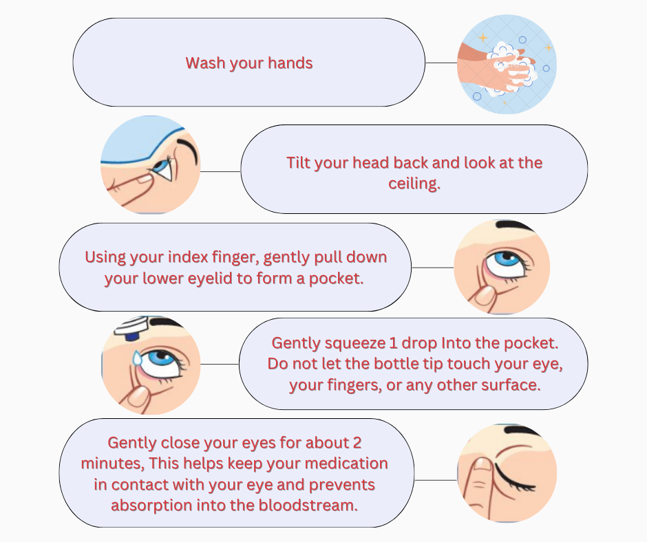
How do I use my eye drops?
Treatment
 🔍 Click to enlarge
Glaucoma Diagnosis
 🔍 Click to enlarge
Glaucoma Reasons
 🔍 Click to enlarge
About Glaucoma
 🔍 Click to read infographic
Glaucoma
By
Dr. Deepti Kulkarni
Consultant & Vitreo Retinal Surgeon
Dr. Anil Kulkarni Eye Hospital, Miraj
ALL WORK AND NO PLAY DOES MAKE JACK A DULL BOY!
The importance of playgrounds and games for children can never be more important than now! With an increasing number of gadgets overpowering our daily routines, outdoor activities need to be given MUCH more precedence than they are getting!
There is growing evidence from both human and animal studies of refractive error showing that ambient light exposure is an important environmental factor involved in the regulation of eye growth.
Documented seasonal variations in eye growth and refractive error progression in childhood (with slower eye growth seen in summer months and faster rates of eye growth in winter months) support a potential role for ambient light exposure in the control of human eye growth. So natural sunlight in the classrooms makes for a healthy learning environment.
Indian population: 13.1% of school children have myopia (near-sightedness or minus glass power). There is a positive association of myopia with studying in private schools vs. government schools, positive family history, higher socio-economic status, studying/reading > 5 hours per day, watching television > 2 hours/day and playing computer/video/mobile games.
An inverse association with outdoor activities/playing is observed with children playing > 2 hours a day. This translates to a simple understanding that 2 hours of outdoor play every day will help us overpower this silent epidemic that we are facing.
SO LET THE CHILDREN PLAY OUTSIDE. A LITTLE SUN AND TAN WILL DO NO HARM!
Written By
Dr. Deepti Kulkarni
Vitreo Retinal Surgeon
Dr. Anil Kulkarni Eye Hospital

Treatment of Cataract
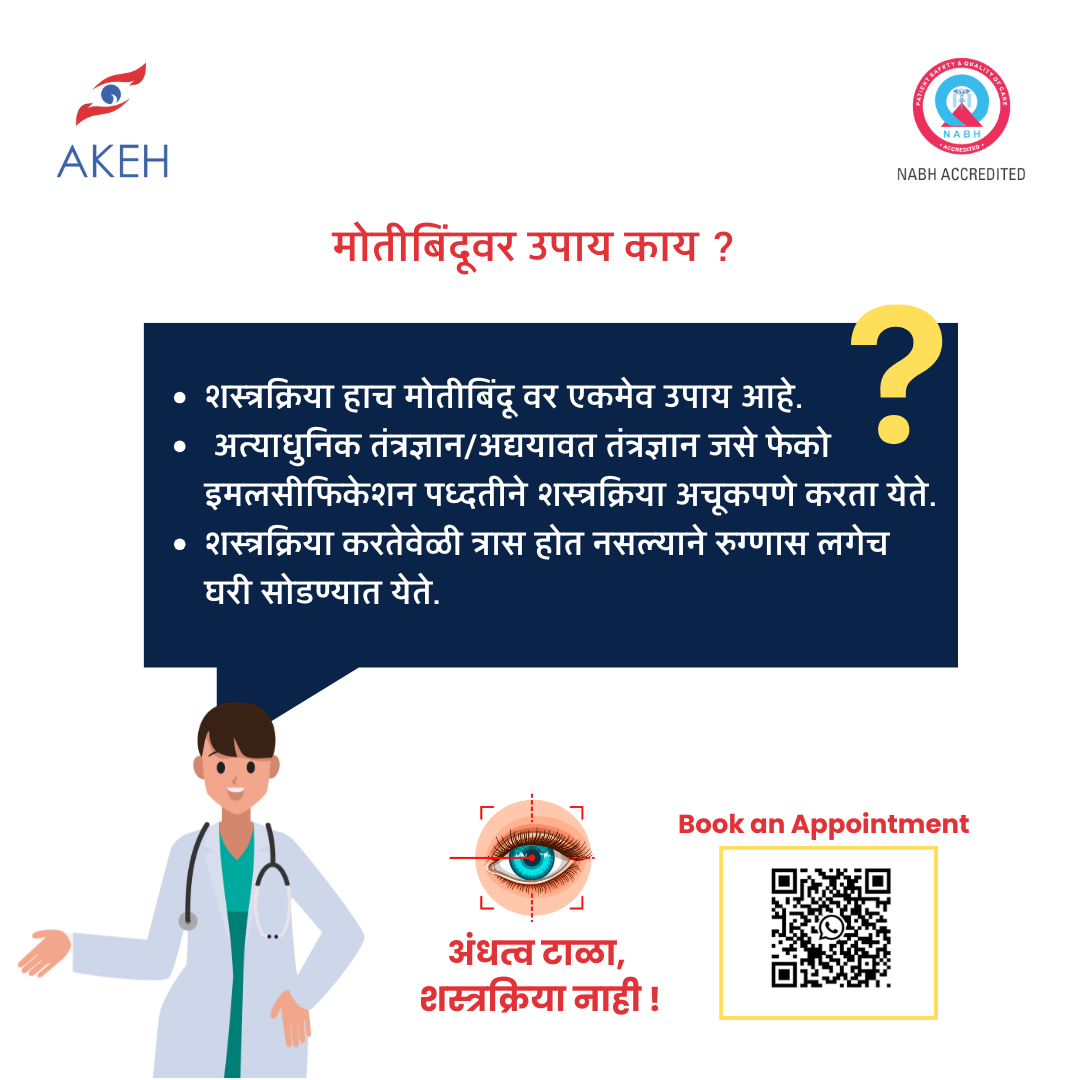 🔍 Click to view full resolution
Cataract Symptoms
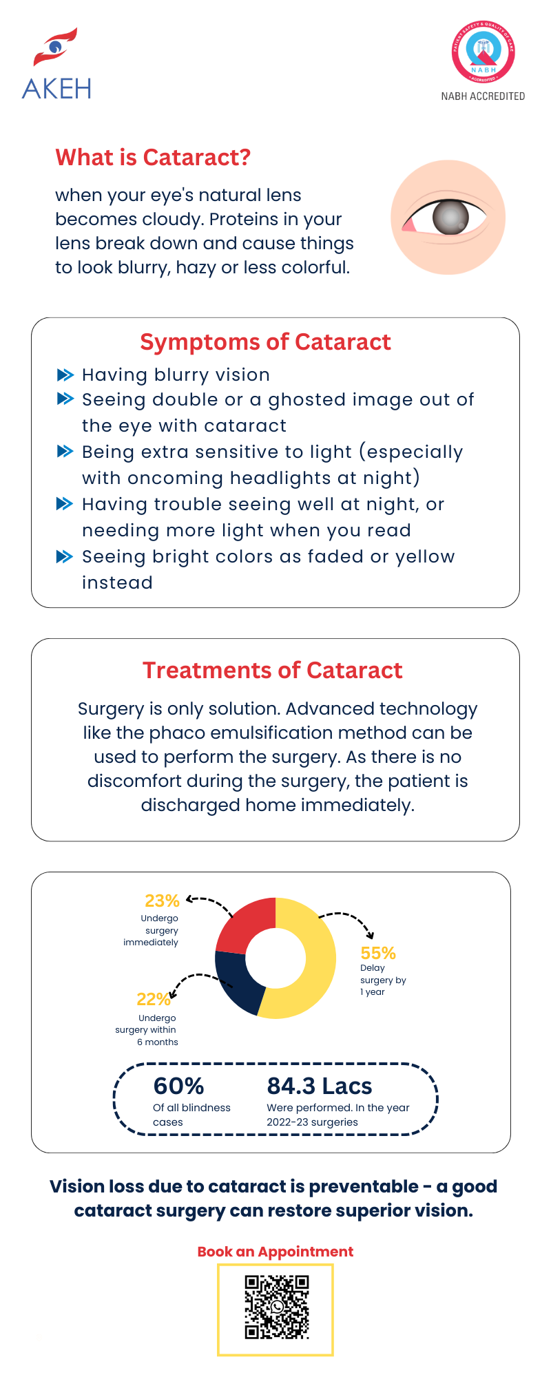 🔍 Click to view full resolution
Cataract Surgery
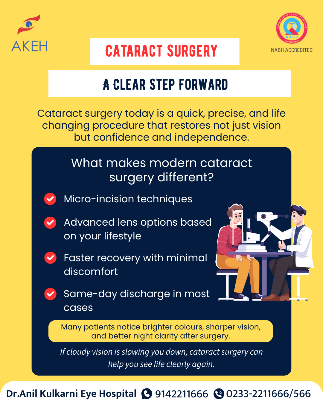 🔍 Click to view full resolution
Cataract Awareness
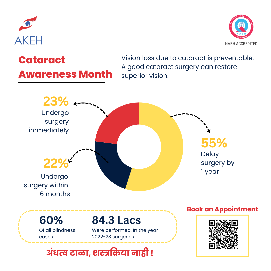 🔍 Click to view full resolution
What is an IOL & How Does It Work
Open in new tabWhat is Cataract & Symptoms
Open in new tabGuide to Cataract Surgery
Open in new tabMyopia
What is Myopia epidemic?
Age Related Macular Degeneration
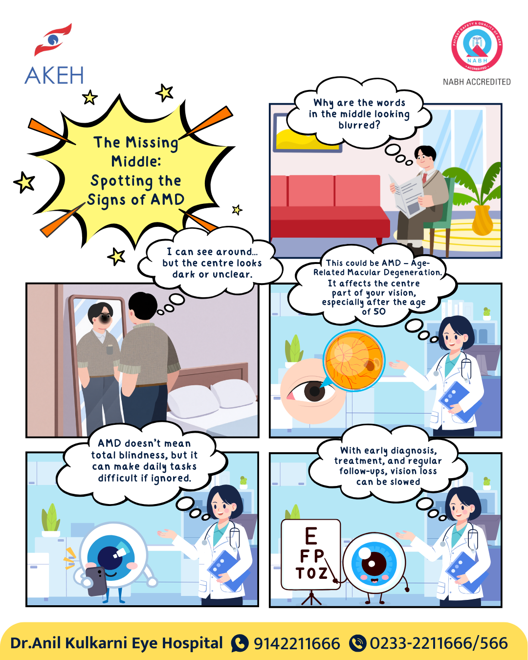
🔍 Click to enlarge
Macular Degeneration Symptoms

🔍 Click to enlarge
Macular Degeneration
Macular Degeneration
Macular Degeneration(Marathi)
Eye Care During Exam
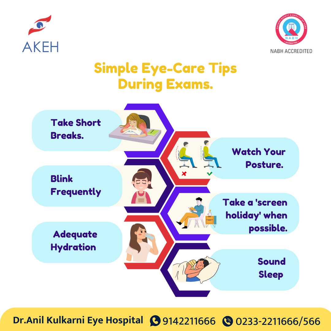 🔍 Click to enlarge
Childhood Diabetic Symptoms
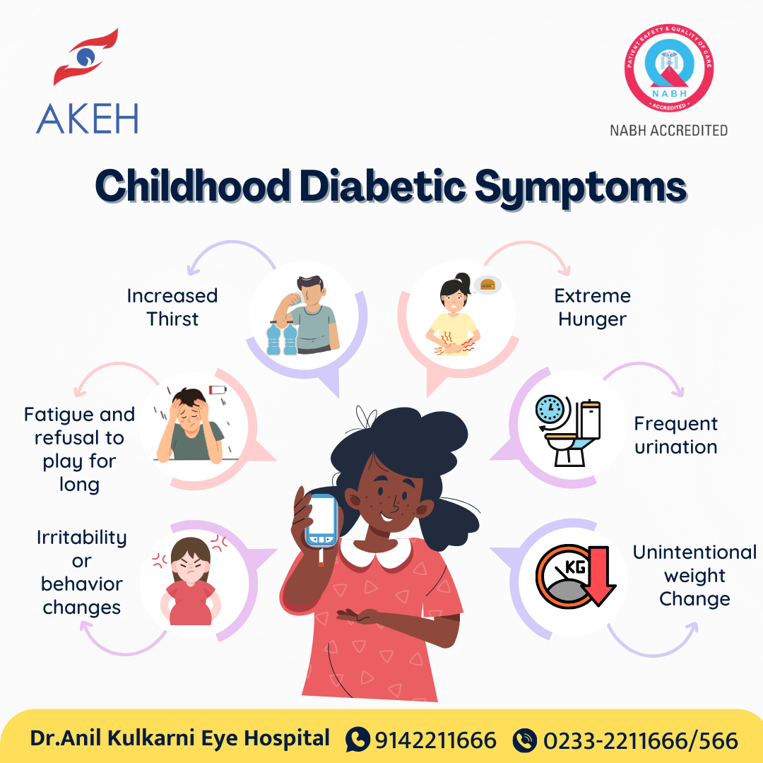 🔍 Click to enlarge
Newborn Baby Eye Care
Know if your children has Glasses
Choosing the Right Frame
Diabetic Retinopathy
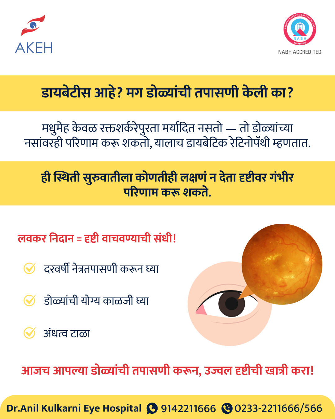 🔍 Click to enlarge
Diabetic Retinopathy
Open in new tabWhat is Retinal Detachment?
Open in new tabRetinal Detachment Treatment
Open in new tabDifficult Surgery – Giant Retinal Tear
Open in new tabDoes Surgery help with Squint ?
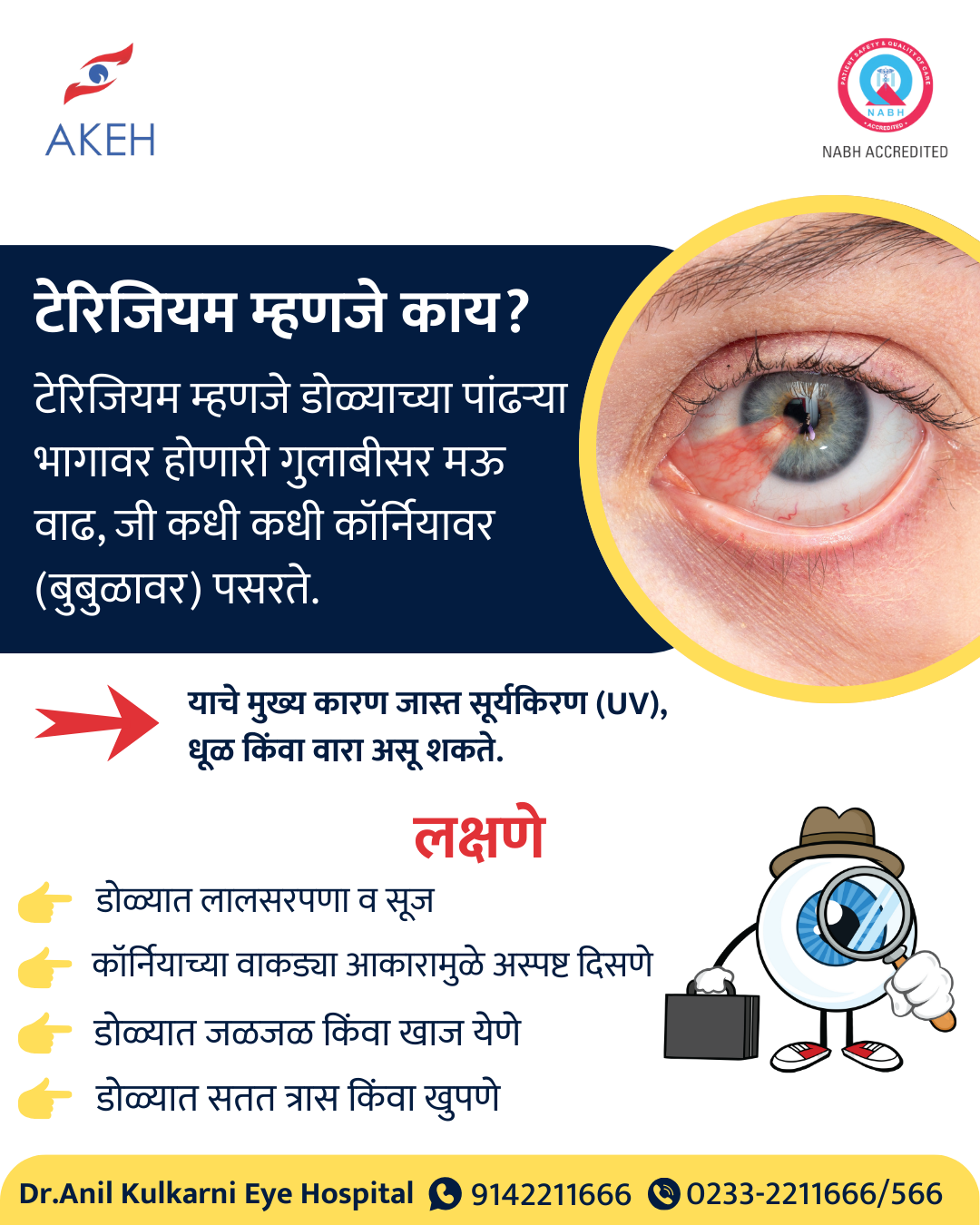
Open in new tabPterygium
Click play to watchPterygium
 🔍 Click to enlarge